Nilotinib Associated With Increased Peripheral Artery Disease Rate in CML
Two studies found higher rates of peripheral artery occlusive disease in nilotinib-treated chronic myeloid leukemia patients than in imatinib-treated patients.
Both a retrospective cohort analysis and a prospective study found higher rates of peripheral artery occlusive disease (PAOD) among patients with chronic myeloid leukemia (CML) who received nilotinib than in those who received imatinib. Both new studies were published online ahead of print on April 5 in Leukemia.

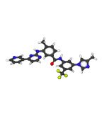
Ball-and-stick model of nilotinib
Previous reports on PAOD and nilotinib treatment in chronic-phase CML patients suggest a frequency of the arterial disorder of between 1.2% and 12.5%. “However, all previously published articles are limited by the description of only clinically manifest PAOD without screening of asymptomatic patients and without prospective monitoring,” wrote authors of the prospective report, led by Theo D. Kim, MD, of the Charit-Universittsmedizin Berlin in Germany. The group prospectively screened all chronic-phase CML patients at one institution between August 2011 and November 2012, using the ankle–brachial index (ABI) and duplex ultrasonography.
The study included 159 patients, some of whom were also included in other CML trials, such as ENESTnd. Of the total cohort, 54 patients were on first-line imatinib; 33 were on first-line nilotinib; 33 had previous imatinib exposure and were on second-line nilotinib; 25 had previous nilotinib and were on another therapy, considered “post-nilotinib”; and 14 were nilotinib-nave patients not receiving imatinib.
ABI was obtained in 129 of the 159 patients (81%), and a pathological ABI indicating PAOD was seen in 24 of 129 (18.6%). There were higher rates of pathological ABI in nilotinib-treated patients than others. Only 6.3% of first-line imatinib patients had pathological ABI, compared with 26% of first-line nilotinib and 35.7% in the second-line nilotinib patients. In the post-nilotinib group, the rate was 16.6%, and 12.5% in the nilotinib-nave group.
Both the first- and second-line nilotinib groups’ rates of pathological ABI were significantly higher than the imatinib group (P = .0297 and .0029, respectively). PAOD that was clinically manifest, defined as “typical peripheral ulcerations or an acute event of PAOD,” were found in five patients, all in the first-line, second-line, or post-nilotinib groups. The relative risk for PAOD defined by ABI was 10.3 for patients on first-line nilotinib vs first-line imatinib, a risk the researchers described as “remarkable.”
The other analysis, a retrospective cohort study, painted a slightly different picture of PAOD and nilotinib, though the increased risk vs imatinib remained clear. That cohort included a huge number of chronic-phase CML patients: 533 with no tyrosine kinase inhibitor (TKI) treatment; 556 treated with nilotinib; and 1,301 treated with imatinib.
The nilotinib-treated patients had an exposure-adjusted risk for PAOD of 0.9 vs the -nave patients, while the imatinib-treated patients had a risk of 0.1 compared with the -nave cohort. A multivariate analysis showed that while nilotinib did not change the PAOD risk vs no , imatinib lowered the risk significantly, and nilotinib was associated with higher overall rates of PAOD than imatinib. The authors, led by Frank Giles, MD, of the National University of Ireland, Galway, concluded that patients initiating therapy for CML should undergo risk factor assessment for PAOD.
Dr. Kim and colleagues also agreed, concluding: “We strongly suggest to capture baseline ABI, biochemical risk factors and to monitor these parameters regularly throughout therapy of CML.”
Between the Lines Podcast: Tazemetostat in Relapsed/Refractory Follicular Lymphoma
November 3rd 2022Expert oncologist/hematologists Bruce Cheson, MD, FACP, and Steven Park, MD, discuss findings from the E7438-G000-101 trial and consider the efficacy of tazemetostat as treatment for relapsed or refractory follicular lymphoma.